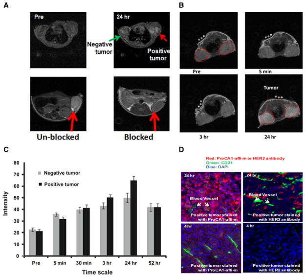
Fig. 3.
a Gradient echo transversal MR images collected prior to injection and at various time points post injection of 3.0 mM ProCA1-affi342-m which is PGEylated ProCA1-affi342 targeting to HER2 in HEPES saline via tail vein. The MRI signal on the positive tumor (SKOV-3, right) exhibits significant enhancement at 24 h post injection. Blocking results confirm the specific binding of ProCA1-affi342 to HER2-positive tumors. b Enhancement changes of MRI intensity in tumors and at 24 h post injection, highest enhancement was observed. c MR imaging of contrast agent ProCA1-affi1907 targeting to EGFR specifically. d Immunostaining show HER2 targeted ProCA1-affi342 has better tumor penetration than HER2 antibodies

