
Figure 8.
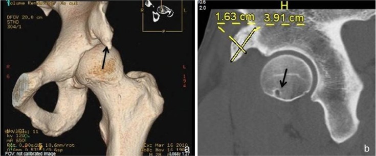
(a) Three-dimensional computed tomography (CT) scan demonstrating prominent anterior inferior iliac spine (AIIS) resulting in subspine impingement and restriction in hip flexion. (b) Sagittal CT image demonstrating AIIS dimensions and point of pathologic contact on the femoral neck (arrow) with terminal flexion and rotation of the hip.