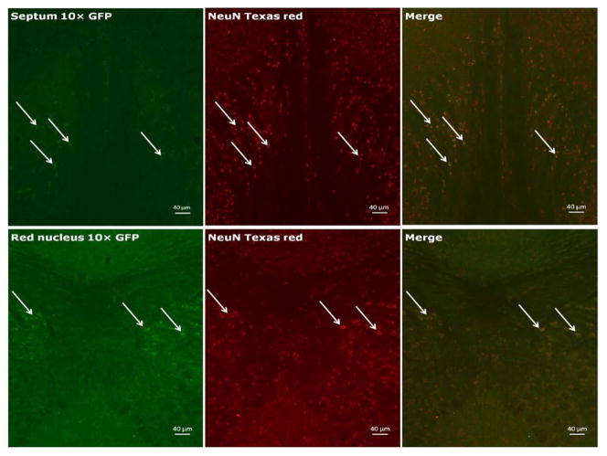
Figure 2.
Representative photomicrographs showing expression of GFP+ cells in the septum (top: most GFP+ cells in the lateral septum and some in the medial septum) and red nucleus (bottom). Arrows label cells positive for GFP (left), the pan-neuronal cell marker NeuN (center), the co-localization of both markers (right).

