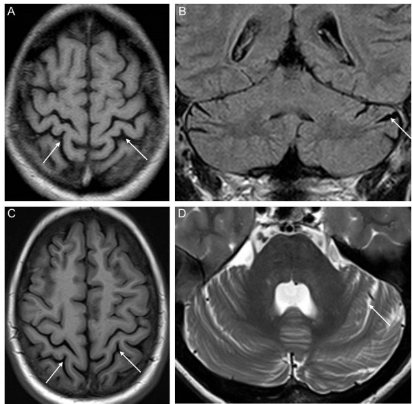
Figure 2.
Brain magnetic resonance imaging (MRI) of the two affected siblings. MRI shows diffuse parietal atrophy (arrows in A, C) and mild cerebellar atrophy (arrows in B, D) in the index patient II-2 at age 31 years (upper row) as well as in his sister II-1 at age 38 years (lower row) (A, C T1 axial; B, FLAIR coronar; D T2 axial).

