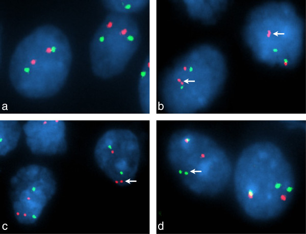
Figure 3.
Interphase fluorescence in situ hybridization analysis of the BRAF locus. FISH probe profiles (a, b, c, BRAF – red; 7p control – green; d, centromeric BRAF – green; telomeric BRAF – red) indicate normal BRAF in GG02 (a) and a classic ‘doublet’ pattern with these probes for duplicated BRAF in GG21 (b, arrows). In GG17 (c, d), FISH preparations indicated a complex alteration; probe profiles showed both duplication of BRAF and a monallelic separation of duplicated BRAF.

