Figure 1.
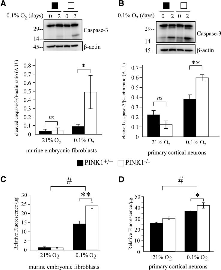
Loss of PINK1 increases vulnerability to hypoxic stress. A, B, Increased caspase-3 cleavage in (A) PINK1−/− MEFs or (B) primary cortical neurons when treated with 0.1% O2 for 2 d. Cell lysates were analyzed by Western blot and quantified for cleaved caspase-3 and β-actin. A representative figure from three independent experiments is shown. C, D, Quantifications from three experiments were pooled for two-way ANOVA analysis. Increased intracellular H2O2 level in (C) PINK1−/− MEFs or (D) primary cortical neurons after exposure to 0.1% O2 for 2 d. H2O2 measurements was performed using Amplex red assay with cell lysates. Samples were normalized to protein concentration and readings from three independent experiments were pooled. *p value < 0.05, **p value < 0.01, #p value <0.001. A.U., arbitrary units.

