
Figure 3.
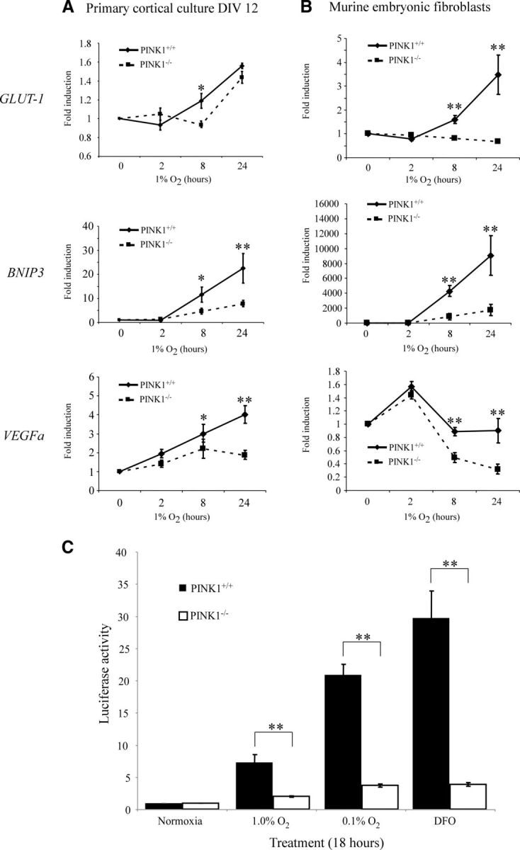
Decreased HIF-1α activity in PINK1−/− cells during hypoxia. A, B, Downstream HRE genes showed decreased induction in (A) PINK1+/+ and PINK1−/− primary cortical cultures and (B) PINK1+/+ and PINK1−/− MEFs when treated with 1% O2 for 0, 2, 8, and 24 h. Quantitative RT-PCR was performed for BNIP3, GLUT-1, and VEGFa and results were normalized to the PPIA signal. The fold induction of each gene was calculated by dividing into the respective starting mRNA quantity at time 0 of each genotype (n = 4–5). C, PINK1−/− MEFs exhibit significantly lowered HRE-promoter activity compared with PINK1+/+ MEFs. Cells were cotransfected with HRE-luciferase and SV40-Renilla constructs and treated with 1% O2 (n = 4), 0.1% O2 (n = 6), and 100 μm DFO (n = 4) for 18 h. HRE-luciferase signal was normalized to the SV40-Renilla signal. *p value < 0.05, **p value < 0.01.