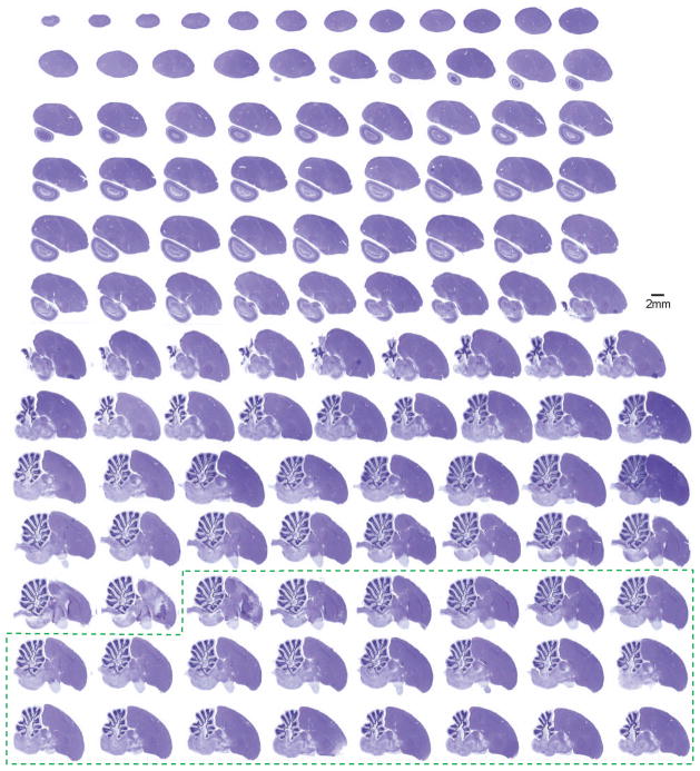
Figure 2. A collection of Nissl-stained zebra finch brain sections presented in the sagittal plane.
The brain was cut into 30 μm thick sections. Alternating sections were stained with either Nissl (Cresyl violet) or myelin (Gallyas silver) stains, respectively, mounted on glass slides, and imaged with an Aperio Scanscope XT. Each 8-bit color image is digitized at high-resolution (0.5 μm/pixel). The entire collection consists of 14 slides of Nissl-stained sections derived from one hemisphere (8–12 sections/slide). Additional sections from the opposite hemisphere are also included with this series (dotted green box), since it is difficult to precisely identify the midline during the course of tissue preparation. Sections from lateral to medial are displayed from left to right, and top to bottom. This series reveals the cytoarchitectonic structure of the zebra finch brain, and complements the myelin-stained sections presented in Figure 3.

