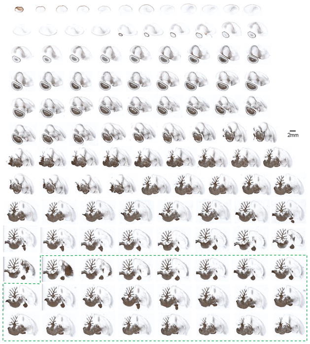
Figure 3. A collection of myelin-stained zebra finch brain sections presented in the sagittal plane.
Sections were obtained from the same brain (in alternating series) as those presented in Figure 2, and are imaged and arranged in an identical fashion. Corresponding myelin and Nissl sections are adjacent to each other and 30 μm apart. This series demonstrates the myeloarchitectonic structure of the songbird brain, and complements the Nissl-stained sections presented in Figure 2. The dotted green box indicates additional sections from the opposite hemisphere.

