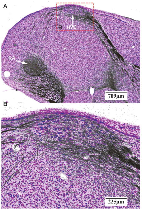
Figure 5. Superimposition and registration of adjacent Nissl and myelin images.
Images from Figure 4 panels C and D are superimposed to illustrate the complex cyto-and myelo-architecture of song nuclei HVC and RA. The Nissl stain is displayed as the blue/purple component in the image, with the Myelin stain in black. Information from the each of the two staining methods complements each other to provide a more complete description of the anatomical structure and connectivity. For anatomical abbreviations, see list.

