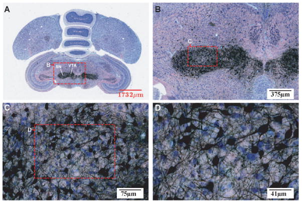
Figure 6. A Tyrosine Hydroxylase (TH) stained section in the transverse plane at progressively higher magnification levels.
Antibodies were applied against TH, which is a limiting enzyme in the catecholamine synthesis pathway, and sections were counter-stained with Giemsa. Neurons synthesizing catecholamines were consequently labeled in black, while others were stained in blue due to Giemsa staining. The ROI (red box) of each image is magnified in the next panel. The immunohistochemical technique complements the Nissl and myelin staining by probing additional organizational characteristics (e.g. common neurotransmitters) of groups of neurons. For abbreviations, see list.

