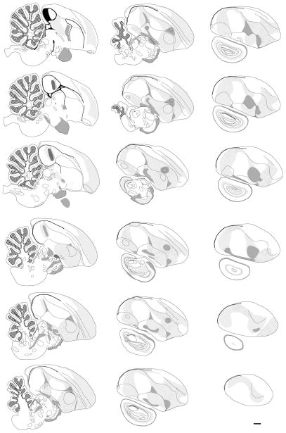
Figure 8. Series of 18 line drawings in the sagittal plane that cover most of the major medial and lateral structures in the zebra finch brain.
Solid lines indicate major contours, brain nuclei, and laminae that can be defined by cytoarchitectonic features in Nissl. Structures and fields shaded in light grey represent white matter fiber fields that are only visible in the myelin-stained material. The orientation and position of fasciculated fiber bundles within these fields are indicated by the thin lines. Structures and fields shaded in dark grey represent commissures and/or thick fiber tracks that are oriented perpendicular to the plane of section. The granule cell layer of the cerebellum is indicated by hatching pattern on a white background; the ventricle is represented as a filled (black) structure. Scale bar = 1 mm.

