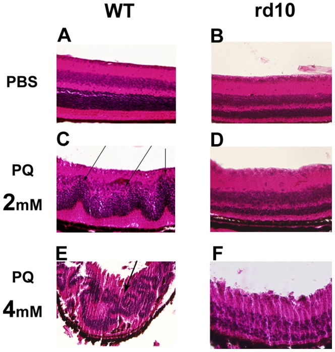
Figure 3. H&E staining of formalin-fixed paraffin-embedded retina sections from rd10 and WT mice injected with 1 µl of 2 mM or 4 mM PQ or PBS.

Normal retina morphology was seen in WT mice (A), while the retina appeared disrupted in these mice following PQ injection (C, E). Arrow indicates wavy appearance of nuclear layers and photoreceptor inner and outer segments in WT retina injected with PQ. This wavy appearance appear extreme following injection of 4 mM PQ. Retinas of rd10 mice, already undergoing retinal degeneration, are typically thinner, due to loss of photoreceptors (B). PQ injection in rd10 mice was not associated with structural alterations similar to the one observed in WT mice (D, F). GCL = ganglion cell layer, INL = inner nuclear layer, ONL = outer nuclear layer, RPE = retinal pigmented epithelium.
