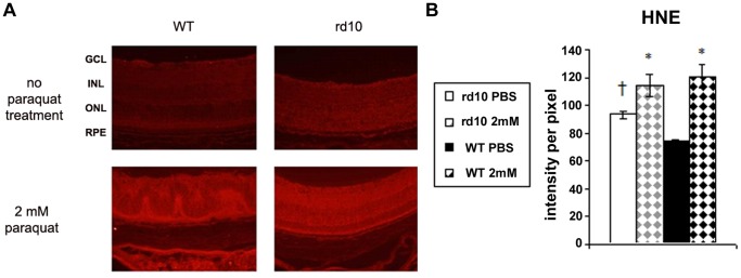
Figure 4. HNE staining of retina sections for assessment of oxidative injury following PQ injection.

Retinas of rd10 and WT mice injected with 1 µl of 2 mM PQ or PBS were labeled with anti-HNE antibody (red; A). GCL = ganglion cell layer, INL = inner nuclear layer, ONL = outer nuclear layer, RPE = retinal pigmented epithelium. (B) Quantification of HNE staining intensity showed marked oxidative injury following PQ injection in WT and the rd10 mice (*p<0.05 as compared to PBS injected eyes of same strain. † p = 0.0002 comparing control (PBS) eyes between the strains; n = 5 in each group).
