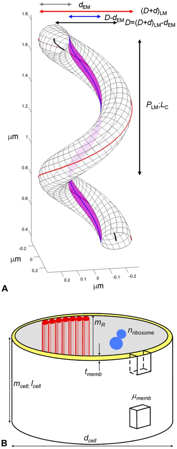
Figure 1. Cell geometry and measurble parameters.

[A] Schematic diagram showing a right-handed, 1.5 turn helical tube with dimensions similar to an average Spiroplasma cell. LM and EM subscripts indicate parameters measured directly by light- and electron microscopy, respectively. one helical repeat (P; vertical black arrow) and corresponding helical centerline, LC–black line, are marked. The basic tube diameter (d; grey horizontal arrow), tube centerline diameter (D;black horizontal arrow), shortest line, LS–blue line–and corresponding inner tube diameter (D-d;blue arrow) and outer coiled-tube diameter (D+d;red arrow) are labeled. The center of the cytoskeletal ribbon, LR, (magenta) follows the geometrically shortest helical line (blue). See Eqs. 2, 17 and 18 for analytical details [14] and Table 1 for actual dimensions. [B] Spiroplasma cell compartments and measured STEM parameters are illustrated. The major mass compartment is a membrane tube of diameter dcell and thickness tmemb (yellow) (see Fig. 7 and Table 1 ) to which a seven-fibered cytoskeletal ribbon (red) is attached. Together these form the dynamic cell envelope. The following parameters were determined on freeze-dried preparations: (i) the mass-per-length of whole, intact cells (mcell); (ii) the mass-per-area of patches of defined areas of empty membrane vesicles (µmemb); and (iii) the mass-per-length of isolated cytoskeletal ribbons (mR) and their component single fibrils (mfibril) [16]. Ribosome counts, nribosome, per unit area of thin sections were used to estimate the number of ribosomes per cell (Eqs. 22, 24).
