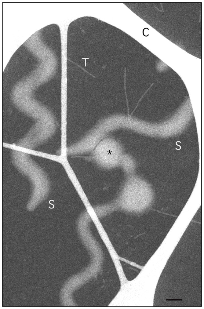
Figure 2. STEM dark field image of freeze-dried, intact Spiroplasma cells.

A thick carbon lace (C) supports a thin (∼3 nm) carbon film. Cells (S) and TMV particles (T), the latter used for mass calibration, are scattered on the carbon support. The cells are polar and have distinct tapered (left edge) or round ends (not seen). Older cells are known to vesiculate, as indicated by the asterisk. Note the difference in mass, as reflected by differences in image brightness, between the straight, uniform tubular cell segments (dimmer) and the heavier (brighter) inflection points of the collapsed coils. Only the straight, uniform segments were used for mass measurements. Scale bar = 200 nm.
