Figure 3.

Esophagogastroduodenoscopy image.
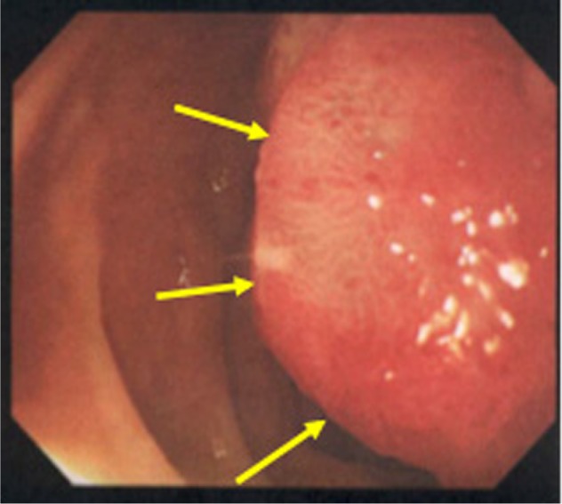
Note: Esophagogastroduodenoscopy reveals a red sessile tumor protruding into the second part of the duodenum (arrows), corresponding to the findings of CT (Figure 1).
Abbreviation: CT, computed tomography.

Esophagogastroduodenoscopy image.
Note: Esophagogastroduodenoscopy reveals a red sessile tumor protruding into the second part of the duodenum (arrows), corresponding to the findings of CT (Figure 1).
Abbreviation: CT, computed tomography.