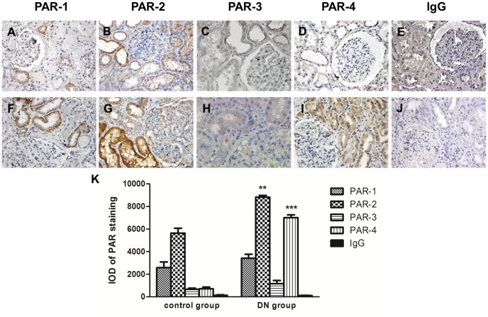
Figure 4. Representative immunohistochemical staining of PAR-1, PAR-2, PAR-3 and PAR-4 in human renal biopsies.
Expression of PARs was demonstrated in renal tissue from patients with biopsy proven diabetic nephropathy (F–I) and non-diabetic control subjects (A–D). Isotype-matched negative control was shown in diabetic nephropathy section (J) and non-diabetic control (E). Magnification: 400X. Quantitative analysis of PAR staining by Image Pro Plus 6.0 software (K); IOD, Integrated Optical Density. **p<0.01; ***p<0.001 compared with control.

