
Figure 5.
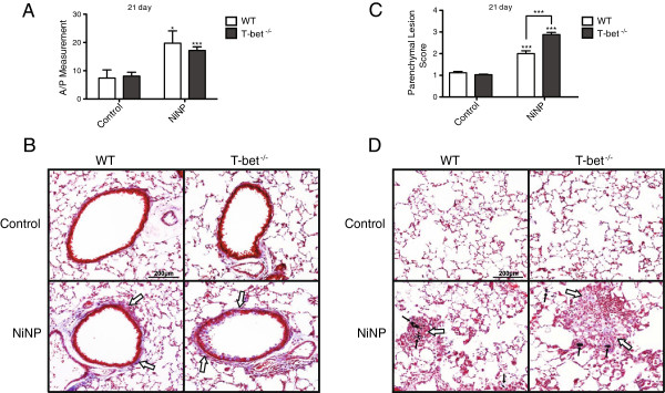
The effects of NiNP on airway fibrosis and parenchymal alveolitis in WT and T-bet-/- mice. A) Cross-sections of airways stained with trichrome were measured for the area to perimeter ratio of collagen deposition. B) Representative images of results from (A) of airways stained with trichrome (open arrows) at low magnification (10×) at 21 days post exposure. C) Lung pathology was then scored for parenchymal lesions in WT and T-bet-/- mice. D) Low magnification (10×) of trichrome stained lung parenchyma sections (open arrows) at 21 days after WT and T-bet-/- mice were initially exposed. Images are a representation of the data shown in (C). Black arrows indicate NiNP aggregates. Data are the mean values ± SEM (n = 5–7 animals/group). *p < 0.05, ***p < 0.001 compared to the control group of the same genotype or as indicated.