
Fig. 1.
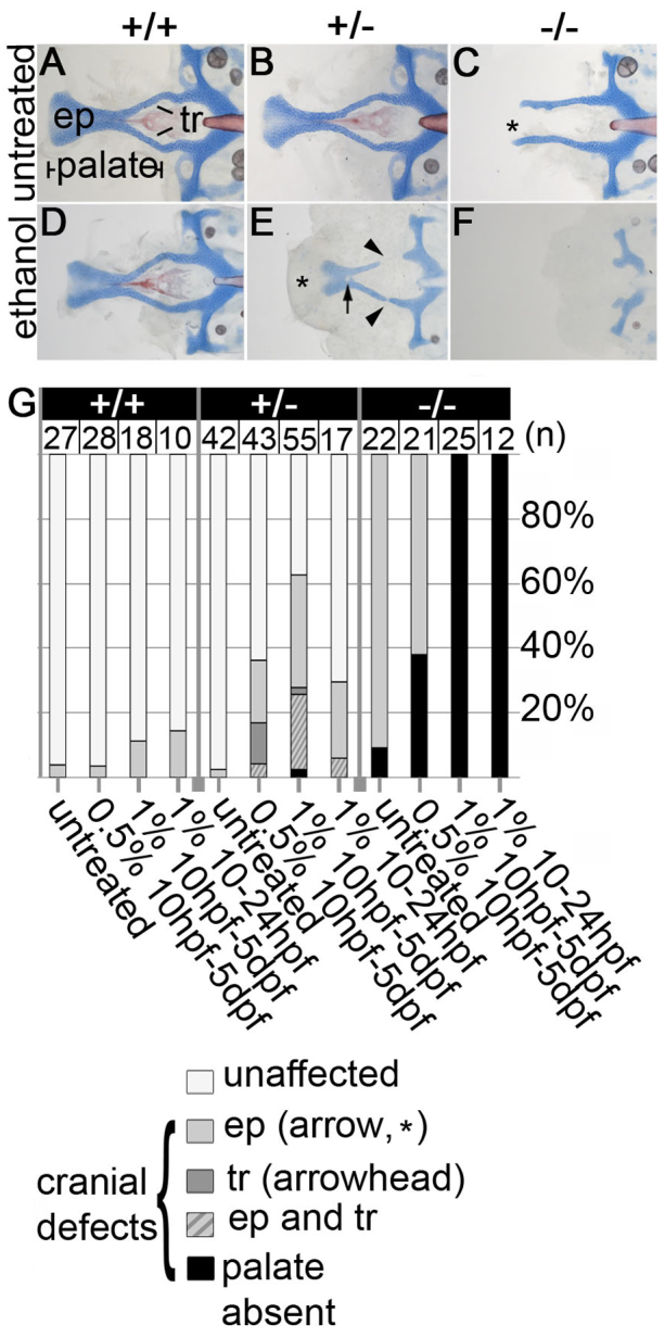
Ethanol exacerbates pdgfra mutant neurocranial defects and reveals haploinsufficiency. (A-F) Flat-mounted 5 dpf zebrafish neurocrania. Anterior is towards the left. (A,B) Untreated wild types and pdgfra heterozygotes develop normally. (C) Untreated pdgfra mutants have clefting of the ethmoid plate (asterisk), although the trabeculae are typically present. (D,E) Neurocrania of embryos treated with 1.0% ethanol from 10 hours post-fertilization (hpf) to 5 days post-fertilization (dpf). (D) Wild-type embryos are predominantly normal following ethanol treatment. (E) Ethanol-treated pdgfra heterozygotes display variable palatal defects, including partial clefting of the ethmoid plate (asterisk), holes in the ethmoid plate (arrow) and breaks in the trabeculae (arrowheads). (F) Ethanol-exposed mutants have an invariant and complete loss of the palatal skeleton. (G) Quantification of palatal defects across genotypes and treatments. ep, ethmoid plate; tr, trabeculae.