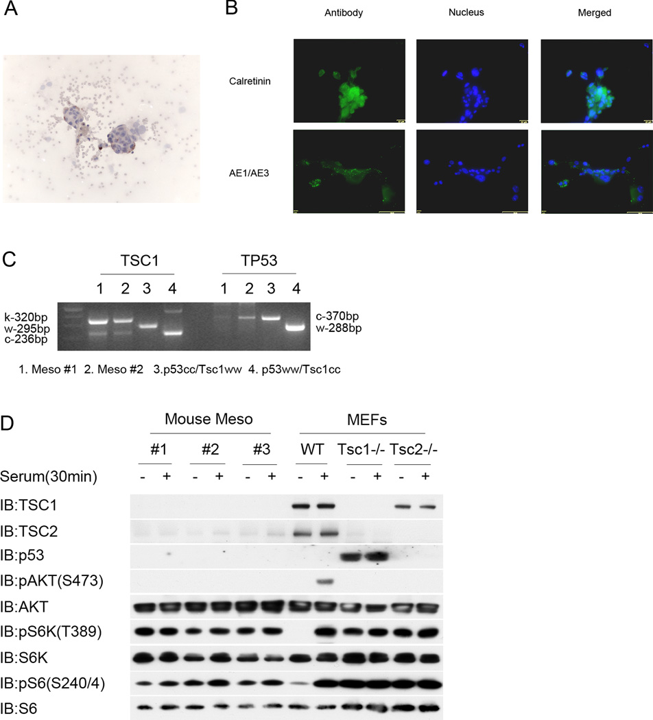
Figure 3. Analysis of mesothelioma cell lines generated from malignant ascites of Cre-treated Tsc1ccTp53cc mice.
A. Cytology preparations demonstrate mesothelioma cell clusters in malignant ascites of Cre-treated Tsc1ccTp53cc mice. Cytospins were stained for pS6(S235/S236) (brown), and counterstained with hematoxylin (blue).
B. Expression of Calretinin and AE1/AE3 by mouse mesothelioma cell lines generated from ascites.
C. Genotyping at the Tsc1 and Tp53 loci on two mouse mesothelioma cell lines and two control DNA samples. Note predominance of the k or knockout allele in the Tsc1 genotyping, and marked reduction in the conditional allele in the Tp53 genotyping, for each of the mesothelioma cell lines, indicative of recombination at both genes.
D. Immunoblot analysis of 3 mouse mesothelioma cell lines in comparison to widltype (WT), Tsc1−/−, or Tsc2−/− murine embryo fibroblast (MEF) cell lines after 48 h of serum starvation (−) or 30 min after serum addback following serum starvation (+). Note absence of Tsc1 and Tp53 in the mesothelioma cell lines; reduced expression of Tsc2; persistent high levels of pS6K(T389) and pS6(S240/S244) without serum; and reduced pAKT(S473) levels following serum addback for the mesothelioma lines. The WT MEF line shows a normal signaling pattern after serum addback.

