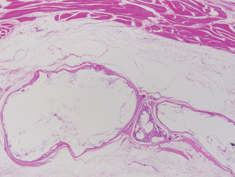
Figure 2.

Poly(D/L)lactide acid plate at 3 months postimplantation. The implant demonstrated fragmentation and giant cell infiltration into the implant. Surrounding loose connective tissue is normal in appearance without evidence of inflammation, cellulitis, or abscess formation. (light microscopy, hematoxylin and eosin stain, original magnification ×40).
