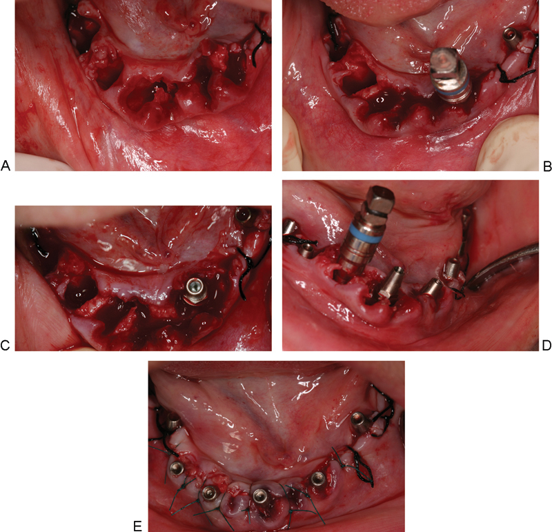
Figure 5.

(A) Atraumatic extraction of the six anterior teeth followed by curettage of the sockets and removing any bony spicules using bone rongeur. (B) Drilling 3 mm in the depth of the lingual wall using pilot drill followed by threaded bone expander no. 2 to allow for bone condensation against the walls of the osteotomy, which aids in better implant stability and positioning the implant buccally without compromising esthetics. (C) MRT implant in place. (D) Continue the same procedure in other sockets. (E) Four postextraction MRT implants and positioning back the flap.
