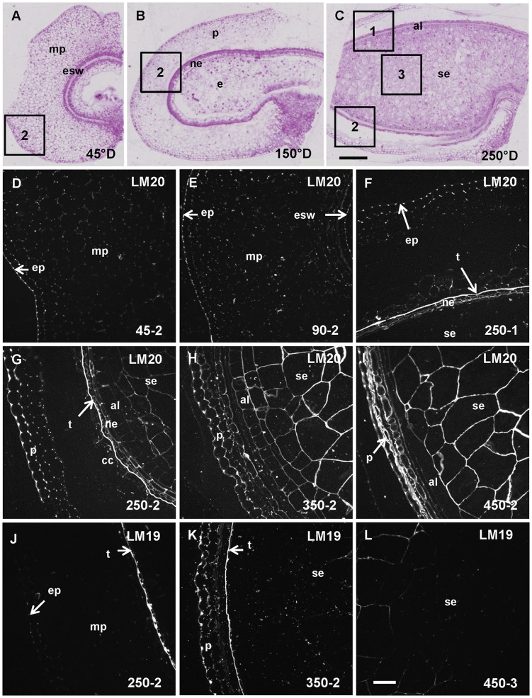
Figure 3. Homogalacturonan (LM20 and LM19 epitopes) deposition in developing wheat grain.
A, B, and C. Half wheat grain harvested at 45°D, 150°D and 250°D stained with toluidine blue showing the evolution of the grain tissues and the regions 1, 2 and 3 where the immunofluorescence acquisitions were taken. D, E, F, G, H, I, J, K and L. Immunofluorescence images localizing methylesterified HG epitope using LM20 and low (or non) methylesterified HG epitope using LM19 antibody. The sections were all treated with lichenase and xylanase prior to immunolabeling. D. LM20 labeling is observed from 45°D in the pericarp and especially in the epiderm of the pericarp (ep). F, G. LM20 labeling is observed only at 250°D in the testa (t), and from 250°D in the endosperm (G, H, I). J. LM19 labeling is observed from 250°D especially in the testa, the pericarp is labeled from 250°D and the starchy endosperm (se) from 350°D (K). al: aleurone layer, cc: cross cells, e: endosperm, esw: embryo sac wall, mp: mesocarp, ne: nucellar epidermis, op: outer pericarp, p: pericarp, se: starchy endosperm. Bars represent 250 µm for A, B and C and 50 µm for the remaining images (all at the same scale).

