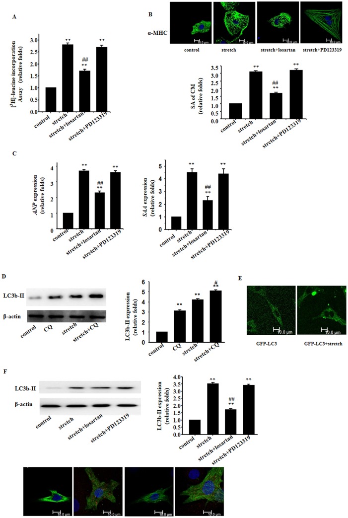
Figure 1. Involvement of AT1 receptor in mechanical stretch-induced cardiomyocyte hypertrophic and autophagic responses.
Cultured cardiomyocytes of neonatal rats were treated with vehicle (Control), stretch in the absence or presence of Losartan (10−6 mol/L) or PD123319 (10−6 mol/L); A: [3H]-Leucine incorporation in cardiomyocytes; B: Cardiomyocyte morphology and size; cardiomyocytes were subjected to immunofluorescent staining for α-MHC (green) and DAPI; Representative photographs were shown from 3 independent experiments (scale bar: 10 µm); surface area (SA) of cardiomyocytes was evaluated by measuring 100 cardiomyocytes from each dish; C: Expressions of Anp and Saa genes evaluated by real time RT- PCR; β-Actin serving as the internal control; D: Western blot analyses for LC3b-II using the anti LC3b-II antibody; β-actin in whole cell lysate was used as the loading control; E: Fluorescence microscopy analysis for GFP-LC3; F: Western blot analyses and immunofluorescent staining for LC3b-II using the anti LC3b-II antibody (red); β-actin in whole cell lysate was used as the loading control. Representative photograms from 3 independent experiments are shown. All data are expressed as mean ± S.E.M from 3 independent experiments, ** p<0.01 versus cardiomyocytes in the control; ## p<0.01 versus cardiomyocytes with stretch.

