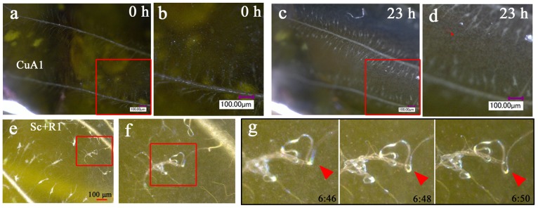
Figure 4. Behavior of tracheal branches and hemocytes.

(a, b) Bright-field image of compartment CuA1 immediately after pupation (0 h postpupation). The boxed region is magnified in (b). Many tracheal branches are already observed at this point, but they are most likely immature and cannot be seen clearly under our observation conditions. They also do not move vigorously. (c, d) The identical CuA1 region 23 h postpupation. Many tracheal branches are observed as white crinoid-like objects, with one side attached to the major trachea. They move relatively vigorously. A single branch exhibits a white knob, from which many thin branches (i.e., tracheoles) radiate. Also note the free-moving hemocytes (indicated by a red arrowhead). (e-g) Tracheal branches in compartment Sc+R1. The boxed region in (e) is magnified in (f), and the boxed region in (f) is further magnified in (g), showing the dynamics of the branch, knob, and tracheoles. The red arrowheads in (g) indicate the identical position in the images over time. The postpupation time is indicated in (g).
