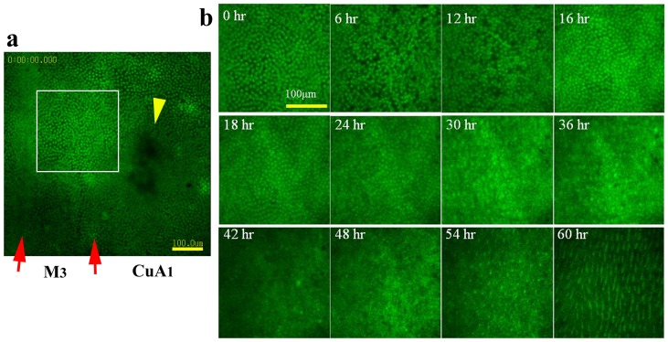
Figure 5. Fluorescent images of array and scale formation.
Wing tissue was stained with CFSE. (a) Stained M3 and CuA1 compartments. Compartment CuA1 exhibits an organizing center, indicated by a yellow arrowhead. The organizing center appears to be resistant to staining. Note that the adjacent M3 compartment does not have this non-stained black area, probably because the M3 compartment does not have an eyespot in adult wings. Also see Figure 6a. Red arrows indicate the major tracheae. The boxed region is enlarged in the subsequent panels. Also refer to Movie S5. (b) Cellular changes over time. At 6 h, the cellular density appears to decrease, and by 16 h the tissue is occupied by densely packed epithelial cells. After 24 h, the cells gradually become arranged, and the wing area increases, which appears to be driven by the contraction pulses that become frequent after 30 h. Scale growth is observed immediately after the cellular row arrangement occurs. All panels are shown at the same magnification.

