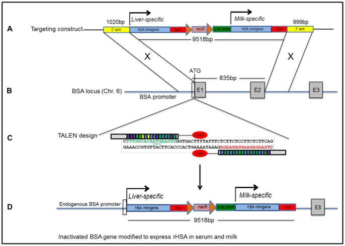
Figure 1. Humanizing the bA locus using TALEN-stimulated HDR.
(A) Structure of the 11.5-kb pHSA-neo targeting construct containing two HSA minigenes and 1 kb targeting arms. The bovine α-lactalbumin promoter/regulatory region is 2 kb in size. Diagram is not drawn to scale. (B) Structure of the bA gene on chromosome 6. Targeting arms are homologous to a region immediately upstream of the endogenous 5′translation initiation codon and within intron 2 (C) Binding of the left and right TALENs. TALE repeats are color-coded to represent each of four repeat variable di-residues (RVDs); each RVD recognizes one corresponding DNA base (NI = A, NG = T, HD = C, NN = G). DSBs are expected to be produced in the 15-bp spacer region in order to stimulate HDR. (D) Proper recombination will result in the deletion of an 835-bp region of DNA, removing bovine exons 1, intron 1, and exon 2, thereby disrupting endogenous bA gene expression and replacing the 5′coding region of the bA gene with two copies of an HSA minigene. HSA1 will be driven by the endogenous bA promoter and will direct rHSA expression to the liver; HSA2 will be expressed in the milk under the α-lactalbumin promoter/regulatory region.

