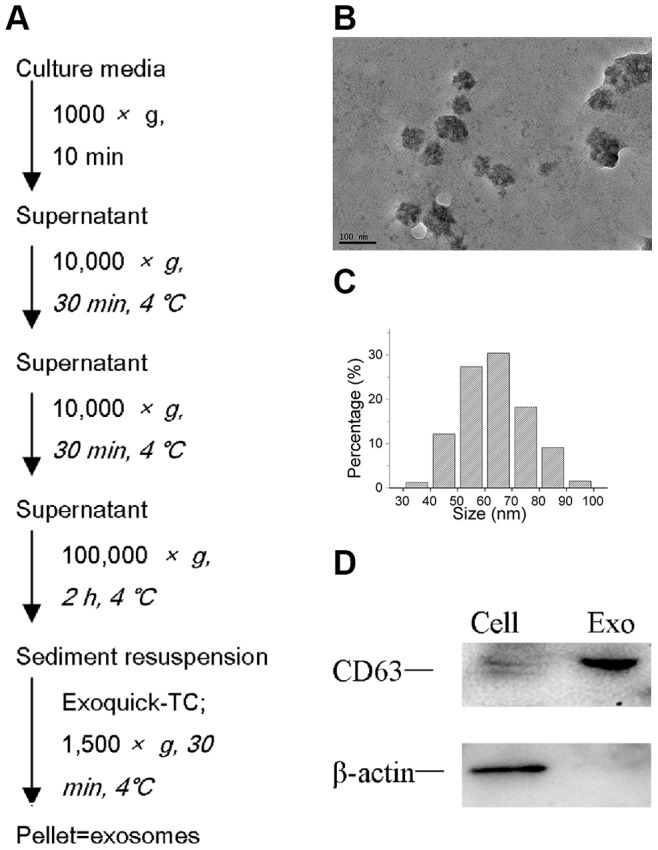
Figure 1. Isolation and characterization of exosomes.

(A) Exosome isolation method used in this study. (B, C) Transmission electron microscopic image of the exosomes. Exosomes are small vesicles measuring from 30nm to 100 nm in diameter. (D) Western blot of CD63 and β-actin in exosomes and cells.
