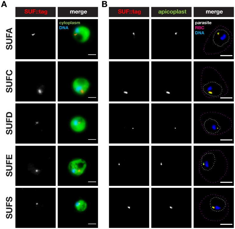
Figure 5. Localization of Plasmodium berghei SUFs to the apicoplast in blood stages.
(A) Epifluorescent micrographs of live suf::tag parasite-infected mouse erythrocytes. Shown are representative micrographs of trophozoite stage parasites. The parasite cytoplasm is labeled by GFP and parasite nuclei by the DNA-dye Hoechst. Bars, 2 µm. (B) Co-staining of fixed trophozoite stage suf::tag parasites using anti-mCherry antibodies and anti-sera against acyl carrier protein (ACP), a signature protein of the apicoplast. Substantial overlap can be observed between the SUF::tag proteins and the signature apicoplast protein in a small structure, i.e. the apicoplast. The outlines of the parasite and the infected red blood cell (RBC) are indicated by white and magenta dotted lines, respectively. Nuclei were stained with the DNA-dye Hoechst. Bars, 2 µm.

