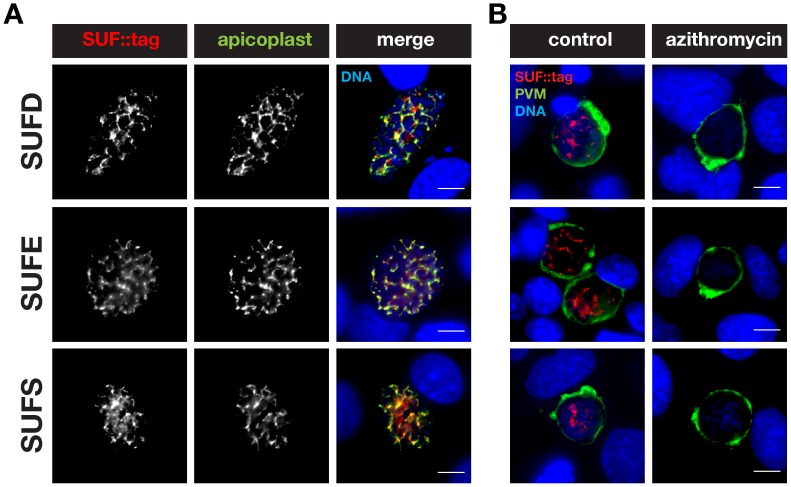
Figure 6. Localization of Plasmodium berghei SUFD, E, and S to the apicoplast in liver stages.
(A) Co-staining of fixed, sufD::tag, sufE::tag, or sufS::tag parasite-infected hepatoma cells 48 h after sporozoite infection using anti-mCherry antibodies and anti-sera against acyl carrier protein (ACP). Note substantial overlap between the SUF::tag proteins and the signature apicoplast protein. (B) Drug treatment of suf::tag-infected hepatoma cells to corroborate apicoplast localization of the SUF::tag proteins. During liver stage development suf::tag-infected cells were left untreated (control) or treated with 1 µM azithromycin. Liver stages were stained with anti-mCherry antibodies and anti-sera against upregulated in infective sporozoite protein 4 (UIS4), a signature protein of the parasitophorous vacuolar membrane (PVM). Nuclei were stained with the DNA-dye Hoechst. Bars, 10 µm.

