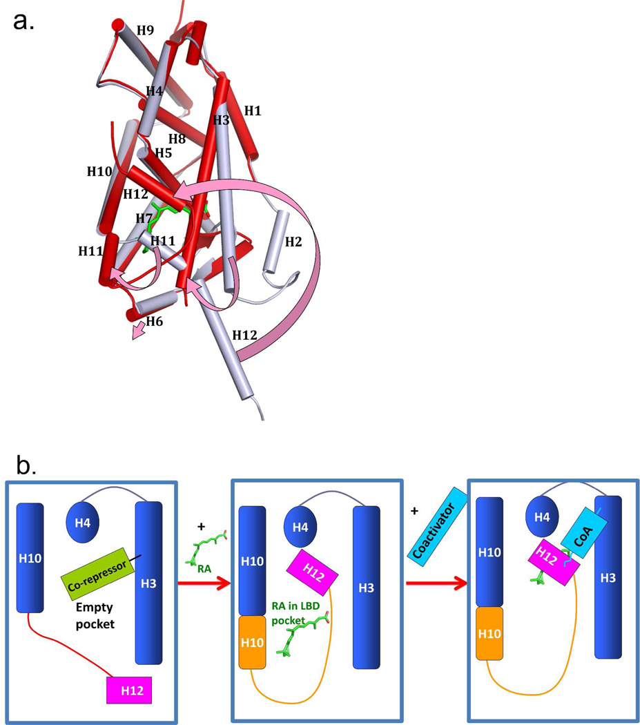
Figure 4.
The ligand induced positioning of helix-12 into the “active conformation”. (A) the superposition of RXRα LBD in the apo-state (grey) and the same receptor in the holo-state (red) with 9-cis RA (green) in the pocket. The four arrows (pink) show the helical movements induced by ligand binding. (B) Illustration of how corepressors, ligand, and coactivators interact with a NR LBD. The blue box represents the LBD portion of NRs. The binding of RA molecules or other endogenous ligands or synthetic agonists results in a major repositioning of helix-12. Ligand binding releases corepressor binding, and causes helix-12 to trap the ligand, by closing on top of its pocket. The resulting active conformation also creates a groove for the binding of coactivator LLxxLL motifs on the surface of the LBD.74 The apo-RXRα is from PBD ID 1LBD, and the 9-cis RA complex is from PDB ID 1FBY.

