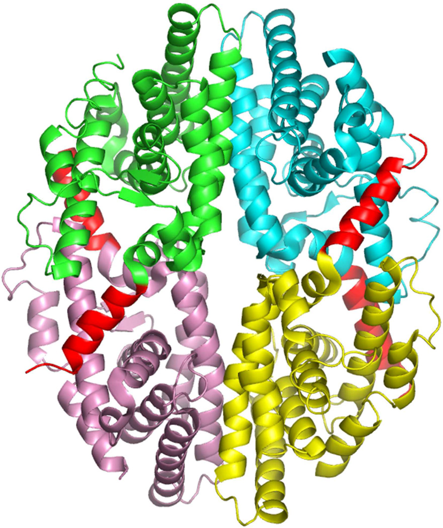
Figure 6.
Structure of the RXRα LBD tetramer in the absence of ligand.233 The four subunits are shown in different colors, but in each case helix-12 is in red. Helix-12 from each subunit traverses to the adjacent subunit to occupy the groove normally reserved for coactivator LLxxLL motif binding. Coordinates are from PDB ID 1G1U.

