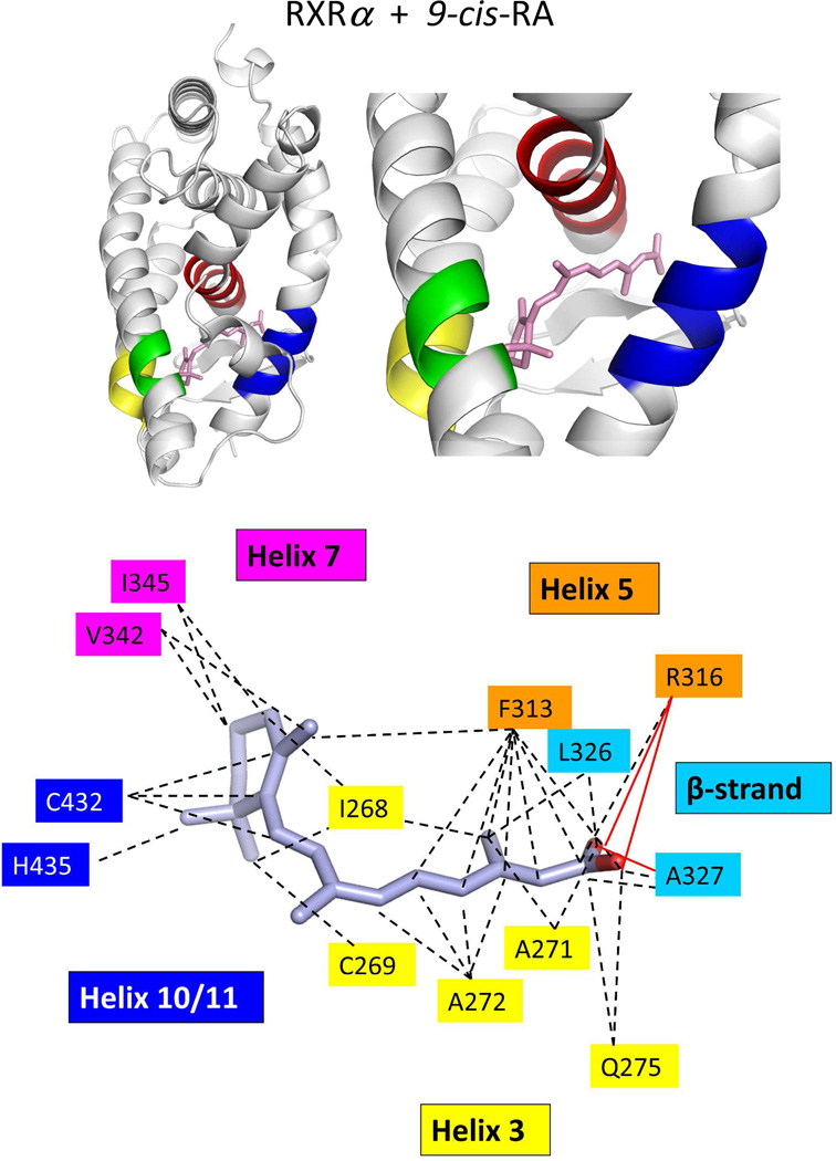
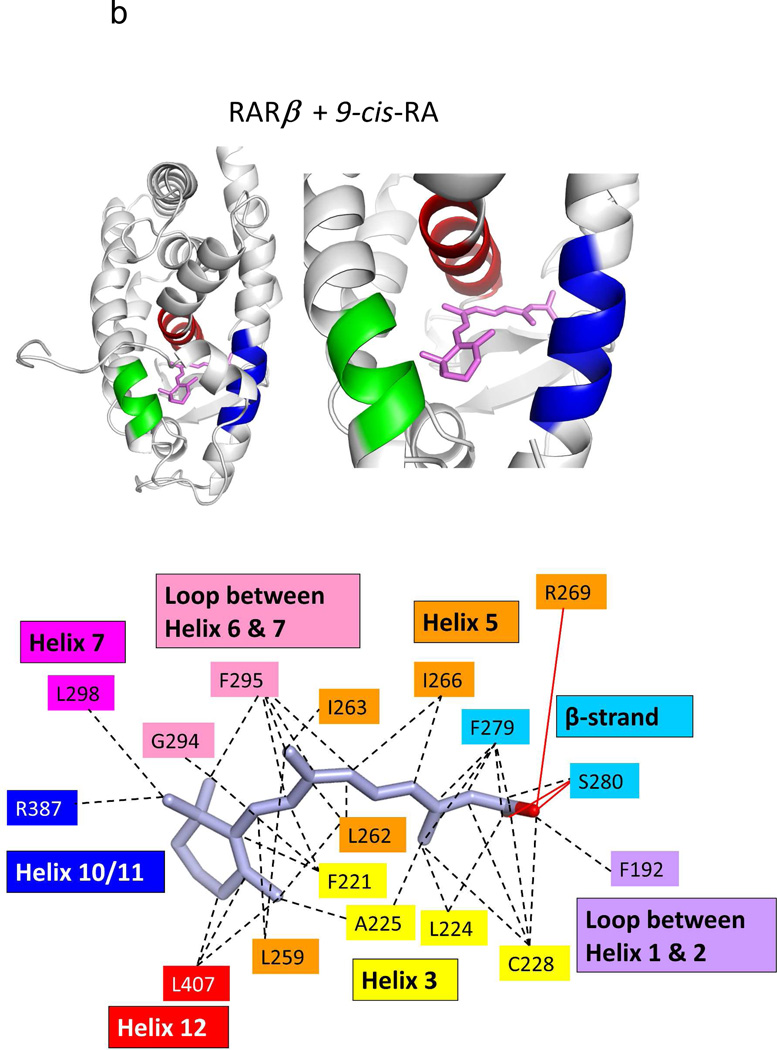
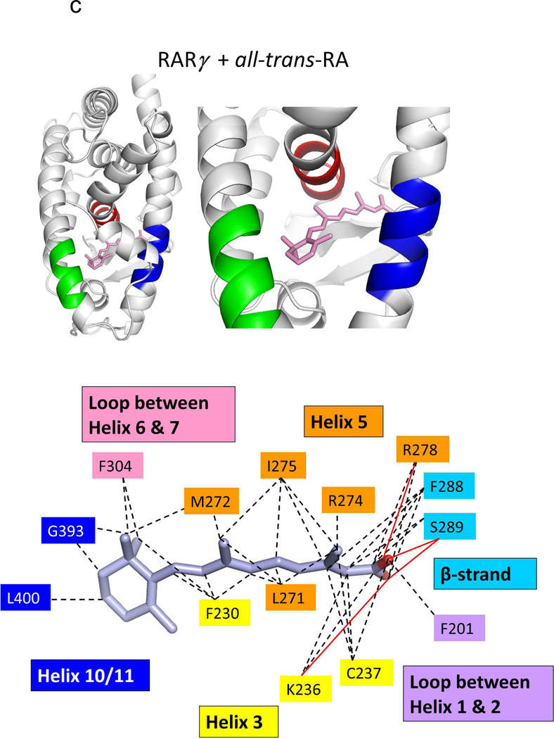
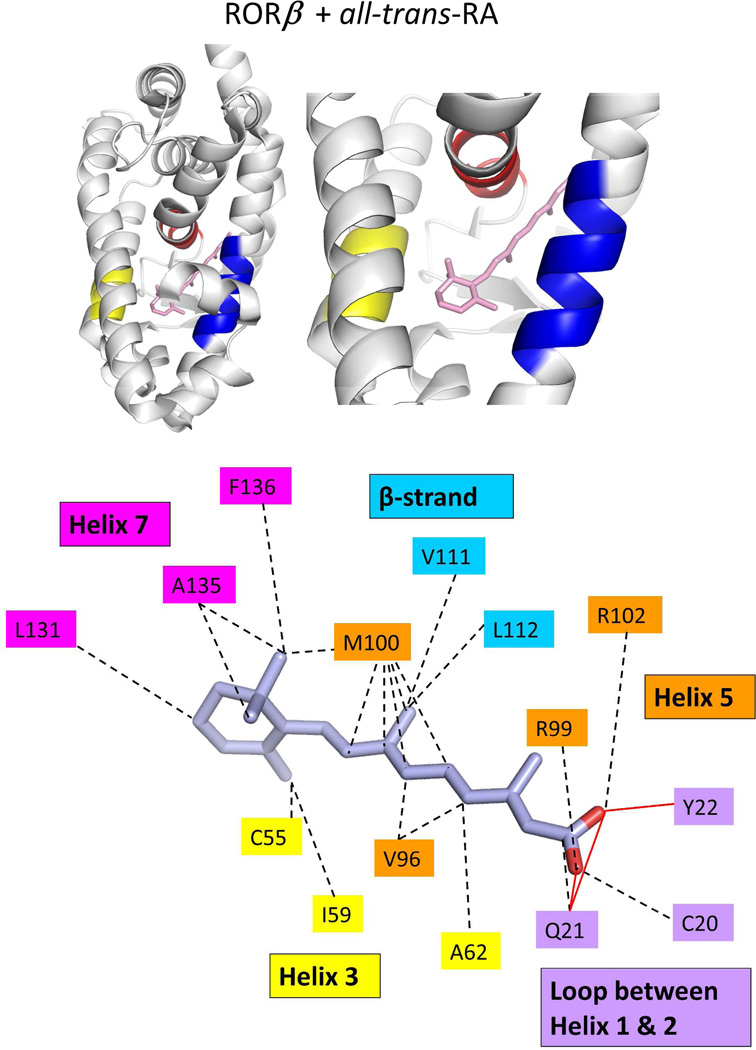
Figure 7.
The binding location and specific interactions of 9-cis RA and all-trans RA inside (A) RXRα, (B) RARβ, (C) RARγ and (D) RORβ LBDs. Dotted lines indicate van der Walls contacts, and solid red lines indicated hydrogen bonding. Coordinates are from PDB ID 1FBY chain A, PDB ID 1XDK chain B, PDB ID 2LBD, and PDB ID 1N4H chain A.


