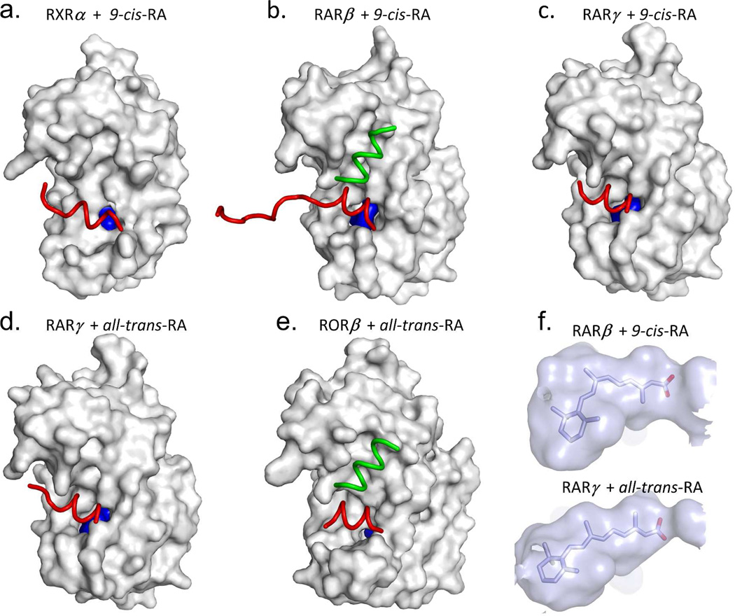
Figure 8.
The degree of ligand burial inside the RAR, RXR and RORβ LBDs. (A–E): The LBDs are in white, the ligands in blue, helix 12 in each case is red, and the coactivator LLXXLL motifs is green. F. The solvent-accessible molecular surfaces from the LBD pockets surrounding 9-cis RA and all-trans RA in RARβ and RARγ, respectively. Coordinates for RARβ+9-cis RA are from PDB ID 1XDK chain B, for RARγ+9-cis RA are from PDB ID 3LBD human, for RARγ+ alltrans RA are from PDB ID 2LBD human, for RXRα+9-cis RA are from PDB ID 1FBY chain A, and for RORβ+ all-trans RA are from PDB ID 1N4H chain A.

