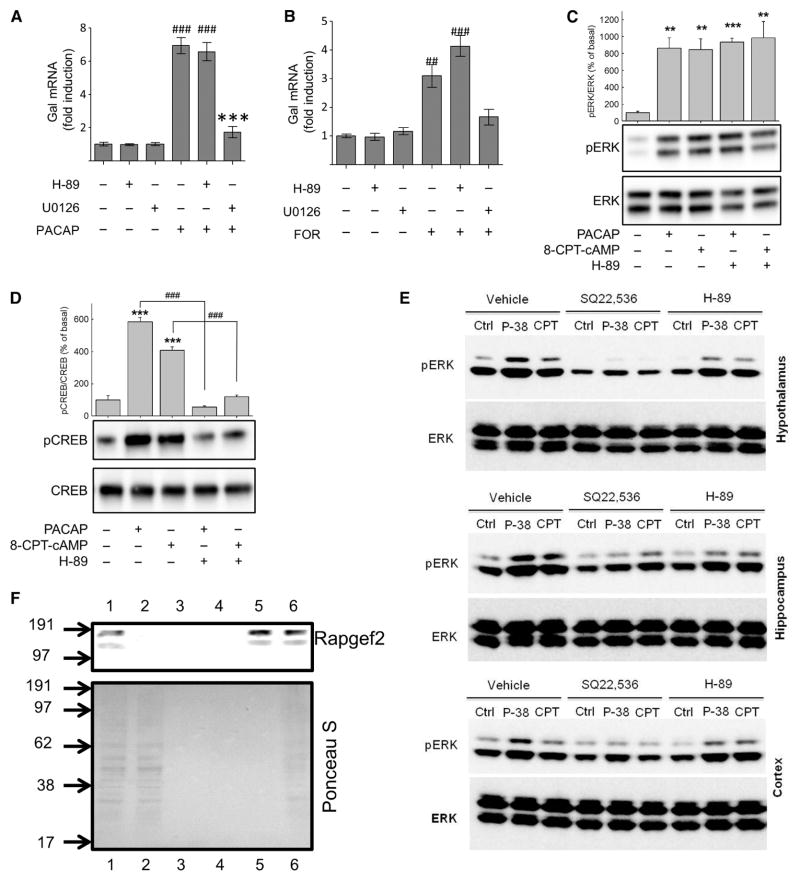
Fig. 1. PACAP and forskolin stimulate galanin expression and ERK activation.
(A and B) Fold induction in Gal mRNA abundance in bovine chromaffin cells (BCCs). Primary cultures of BCCs (n = 3 wells per condition) were treated with (A) 100 nM PACAP-38 or (B) 25 μM forskolin (FOR) for 6 hours. Where indicated, cells were pre-treated with 10 μM H-89 or U0126 for 30 min. RNA was harvested and subjected to quantitative reverse transcription polymerase chain reaction (RT-PCR) analysis. Data are expressed as mean fold induction ± SEM in Gal mRNA abundance compared to that of untreated controls and were analyzed by one-way analysis of variance (ANOVA) with Bonferroni-corrected t tests. ##P < 0.01, ###P < 0.001, when comparing PACAP-38– or forskolin-treated cells with untreated cells; ***P < 0.001, when comparingtreatmentalone and treatment with inhibitors. Experiments represent quadruplicate wells from a single experiment, and experiments were repeated three or more times with similar results. (C and D) Analysis of ERK and CREB phosphorylation. Western blotting analysis of (C) ERK phosphorylation and (D) CREB phosphorylation at Ser133 in BCCs treated with either 100 nM PACAP-38 or 100 μM 8-CPT-cAMP after 10-min pretreatment with 30 μM H-89 or vehicle [0.02% dimethyl sulfoxide (DMSO)]. Data for each experiment (n = 4) were analyzed by one-way ANOVA with post hoc Bonferroni-corrected t tests. **P < 0.01, ***P < 0.001, compared to untreated controls; ###P < 0.001, when comparing cells with or without H-89. Western blots are from a single experiment and are representative of four independent experiments. (E) Analysis of ERK activation in cultures of primary neurons from rat hypothalamus (upper), hippocampus (middle), and cortex (lower). Neurons were pretreated for 30 min with 10 μM H-89, 1 mM SQ22,536, or vehicle (0.02% DMSO) before being treated for 15 min with 100 nM PACAP-38 or 100 μM 8-CPT-cAMP (CPT) or being left untreated (Ctrl). Samples were analyzed by Western blotting with antibodies specific for phosphorylated ERK (pERK) and total ERK. Data are representative of three independent experiments. (F) Lysates from BCCs were subjected to cAMP-agarose affinity purification. Fractions from affinity chromatography were analyzed by Western blotting for Rapgef2 (upper panel), and membranes were stained with Ponceau S (lower panel). Lane 1: crude lysate (1:5 dilution); lane 2: elution with lysis buffer; lane 3: elution with basic wash buffer; lane 4: pooled elutions with high-salt and no-salt wash buffers; lane 5: elution with 50 mM cAMP; lane 6: heat-treated cAMP-agarose resin (prewashed). Numbers to the left of the images represent molecular mass markers (kD).

