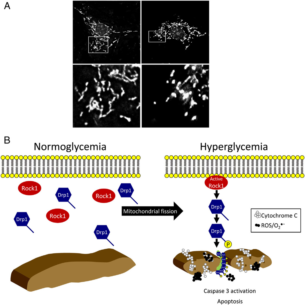
Figure 2.
Mitochondrial fission and fusion. (A) Mitochondrial networks visualized with MitoTracker Red (Life Technologies) fluorescent dye to monitor mitochondrial morphology under (left) normal or (right) high-glucose conditions. Mitochondria appear as long, tubular, and sometimes branched structures that spread throughout the cytoplasm. However, under high-glucose conditions, they appear dense, small, and fragmented. (B) Mitochondrial fission is driven by Drp1, which resides primarily in the cytoplasm. Under hyperglycemic conditions, Drp1 is activated and recruited to the mitochondria. Drp1 then forms spirals around mitochondria at fission sites, which promote the constriction of mitochondria.

