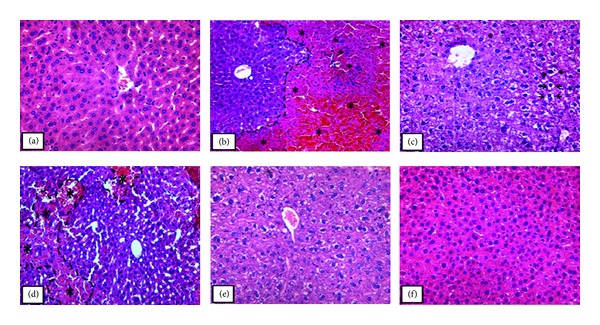
Figure 1.

Photomicrograph of the liver in mice that received orally (a) saline, (b) acetaminophen on last day of treatment (250 mg/kg), (c) silymarin (200 mg/kg), and ((d)–(f)) acetaminophen, after being treated for 7 days with the essential oil of Thymus vulgaris (TEO), 125, 250, and 500 mg/kg, respectively. In (a) the liver showed normal morphology; (b) presence of necrosis and hemorrhagic points (*) in the defined area; ((c) and (e)) parenchyma stands out for having vacuolated hepatocytes (arrows); (d) observed necrotic areas (*); (f) hepatic parenchyma morphology similar to that observed in the control. Original magnification 40x in (a), (c), (e), and (f); original magnification 20x in (b) and (d). The sections stained with hematoxylin and eosin.
