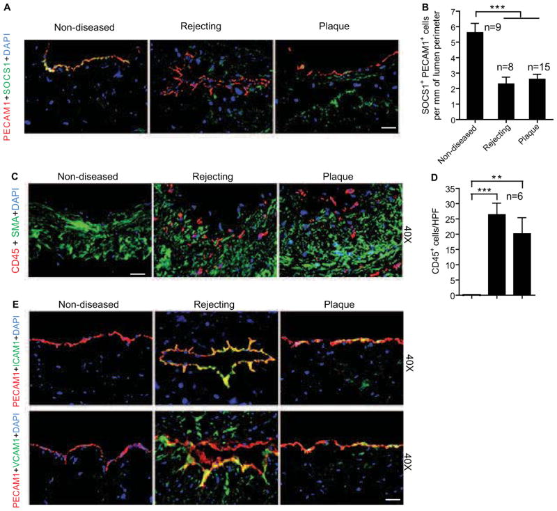
Fig. 1. Loss of SOCS1 expression correlates with clinical pathological vascular remodeling.
Human coronary artery specimens with GA from chronically rejecting heart allografts and with atherosclerotic plaques or no disease from non-transplanted hearts were collected. (A–B) Dramatically decreased endothelial SOCS1 expression in diseased vessel wall. Endothelial SOCS1 expression is demonstrated by immunofluorescence analysis of artery cross-sections stained for SOCS1 and the endothelial marker, PECAM-1 with DAPI labeling of nuclei. Representative images are shown in A with quantifications in B. Scale bar: 50 μm. (C–D) Inflammatory cell and vascular smooth muscle cell accumulation are demonstrated by immunofluorescence analysis of artery cross-sections stained for the leukocyte common marker, CD45 and the smooth muscle cell marker, smooth muscle α-actin (SMA) with DAPI labeling of nuclei. Representative images are shown with quantification in the histogram. Scale bar: 50 μm. (E) Induction of endothelial adhesion molecules has the opposite expression pattern as endothelial SOCS1. Representative histological analysis of artery cross-sections stained with ICAM-1 or VCAM-1 and PECAM-1 antibodies. Immunofluorescence sections were counterstained with DAPI. Scale bar: 50 μm. Data presented in B and D are mean±SEM from independent clinical specimens of varying number per group as indicated; **P<0.01, ***P<0.001, one-way ANOVA followed by Bonferroni’s post-hoc test.

