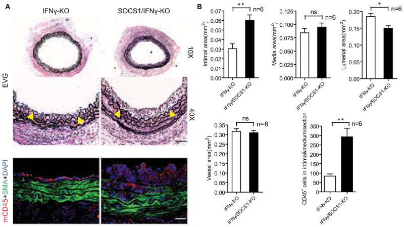
Fig. 2. SOCS1 deletion enhances GA in mice.
Aortas from IFNγ-KO or SOCS1/IFNγ-KO male B6 donors were transplanted to female B6 recipients and the allografts were harvested at 2 weeks post-operatively. (A) Histological analysis of artery grafts by EVG staining and immunostaining by anti-SMA and anti-CD45 antibodies with DAPI labeling of nuclei. Representative photomicrographs are shown. Arrowheads mark the internal elastic lamina to delineate the intima from media. Scale bar: 50 μm. (B) Morphometric assessment of artery graft intima, media, lumen, and vessel areas and quantification of CD45+ cells infiltrating the intima and media of each artery graft cross-section. Data are mean±SEM from 6 mice per group; *P<0.05, **P<0.001, unpaired t-test.

