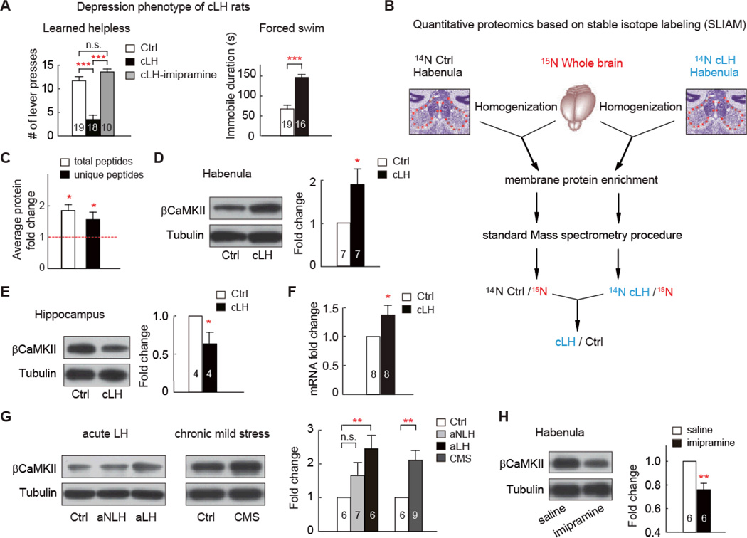
Fig. 1. βCaMKII is upregulated in the LHb of animal models of depression.
(A) Depression phenotypes of cLH rats. Numbers in the bars indicate number of animals used. Note in LH test, maximal number of bar presses is 15. (B) Experimental outline of the high-throughput quantitative proteomics based on stable isotope labeling. Briefly, habenula of unlabeled (14N) WT or cLH rats were dissected, homogenized, and mixed in a 1:1 ratio with total brain homogenate from a 15N-labeled rat. Membrane fraction was enriched, and 100 µg protein sample was used for standard mass spectra analysis. 14N /15N ratio for each identified peptide was calculated. Peptide ratios for each protein were then compared between cLH and control sample. Details see methods. (C) Proteomic analysis of βCaMKII, based either on total peptides, or unique peptides (peptides not shared by other CaMKII family members) identified in 3 independent proteomic runs. (D, E) Western blot analysis showing change of βCaMKII in membrane fraction of habenula (D) or hippocampus (E) of cLH rats. Tissue amounts of tubulin were used as loading control. Protein expression was normalized by control amount. (F) qPCR analysis of βCaMKII mRNA in habenula. (G) βCaMKII level increase in acute learned helpless and chronic mild stress (CMS) depression models. aLH and aNLH were rat groups subjecting to LH stress but did (aLH), or did not (aNLH) display LH symptom. (H) Western blot analysis showing level of βCaMKII in membrane fraction of habenula of cLH rats treated with saline or antidepressant imipramine. Data are mean ± SEM. * p < 0.05 ** p < 0.01, *** p < 0.001 compared to control group, n.s., not significant, two-tailed Student’s t-tests for two-group comparison, one-way ANOVA with Bonferroni post hoc analysis for multiple-group comparison.

