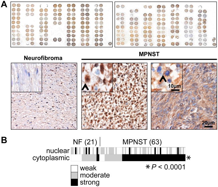
Figure 1. Survivin is highly expressed in human MPNST specimens; cytoplasmic survivin expression is more pronounced in MPNSTs as compared to plexiform neurofibroma.
A. Upper panel depicts the entire TMA stained for survivin; a range of expression levels can be observed. Representative photographs of survivin stained neurofibroma (left) and MPNST spots (middle and right) are shown in the lower panel. Neurofibroma image was captured in 20×, inset demonstrating normal peripheral nerve negative for survivin. MPNST images captured at 20× demonstrating both nuclear (best elucidated in middle picture; inset picture was taken at 40×, arrow marks nuclear staining) and cytoplasmic (best elucidated in right picture; inset picture was taken at 40×, arrow marks cytoplasmic staining) survivin expression (scale bars are included); B. Heat map representation of nuclear and cytoplasmic survivin expression levels in each of the evaluable TMA spots. Cytoplasmic survivin expression was found to be statistically significantly more pronounced in MPNST as compared to neurofibroma.

