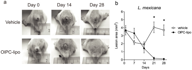
Figure 3. In vivo efficacy of OlPC-liposomes against L. mexicana CL.

Female Balb/c mice received 10 × 106 stationary phase L. mexicana promastigotes at the base of tail. After 12 weeks, mice were treated 2 × 5 days with PBS vehicle or OlPC-liposomes (36–90 μg/day) administered with a tattoo device. Lesions were measured from the start day of the treatment (Day 0) from individual pictures taken on Day 0, 7, 14, 21 and 28 for each mouse. Lesion areas were calculated using the Adobe Photoshop® software. Representative pictures of lesions from each group are shown in (a), while mean lesion sizes (±SEM) up to Day 28 are presented in (b); (*) P < 0.05 between treated and vehicle groups (n = 6).
