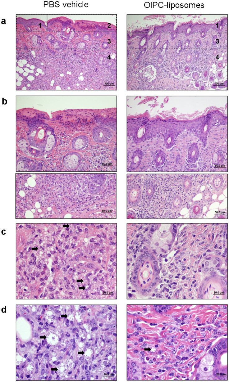
Figure 5. Tattoo-mediated administration of OlPC-liposomes reduces parasite burden and inflammation in the CL lesions induced by L. major and L. mexicana.
On Day 28 following start of treatment, L. major- (a to c) and L. mexicana- (d) infected animals treated by tattooing with PBS vehicle (left panel) or OlPC-liposomes (right panel) were sacrificed and the skin around the lesion was taken and processed for histology using H&E straining. Numbered areas delineate examples of major findings: 1-epidermal hyperplasia, 2-focal ulceration, 3-dermal fibrosis, 4-inflammation. Arrows point representative examples of amastigote-containing histiocytes. Data show representative pictures of 2 independent experiments.

