Figure 7.
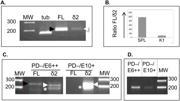
Detecting alternatively-spliced Pax5 isoforms in sorted B cell populations. (A) RT-PCR using RNA from freshly isolated trout K1 or SPL cells. (B) qPCR, ratio fold-change between FL and Δ2 Pax5 isoforms in both SPL and K1. (C) RT-PCR on RNA from fixed, sorted SPL cells (either PD−/E6++ or PD−/E10+) using primers to amplify either FL Pax5 or Δ2. Expected size for FL: 237 bp (black arrow); for Δ2: 219 bp (white arrow). White asterix: non-specific 180 bp band, not Pax5. (D) End-point PCR as in C, but using primers to detect presence of exon 6-containing amplicon, 207 nts. MW, molecular weight (in bps).

