Figure 1.
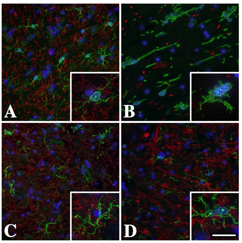
Temporal lobe subcortical white matter microglia/macrophages in male Pelizaeus-Merzbacher disease (PMD) patients are activated and express Iba-1. (A) Iba-1+ (green) resting microglial morphology in the U-fibers from a normal individual who suffered a myocardial infarct. Microglial processes are long, thin and branching (inset). There are abundant myelin basic protein (MBP+) myelinated axons (red) in this region. (B) Activated microglia/macrophages with short thickened processes and amoeboid morphology in the U-fibers from a severely affected PMD patient with a T42I missense mutation in PROTEOLIPID PROTEIN 1 (PLP1). The lack of MBP staining in this section reflects the virtual absence of white matter in this patient. (C) Microglia/macrophage morphology in the U-fibers from this mildly-affected PLP1-null patient is more similar to resting (A) than to activated (B) cells, although there is some thickening of the processes (inset). (D) Microglia/macrophage morphology in the U-fibers from this moderately affected PLP1 gene duplication patient is intermediate between the missense mutation (B) and the PLP1-null patient (C). Scale bar: 50 μm, insets 20 μm.

