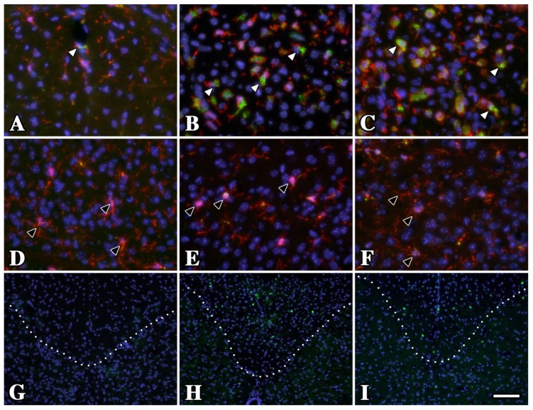
Figure 3.
Oligodendrocytes in gray matter from P16 rsh and msd mice do not undergo a UPR and microglia/macrophages exhibit resting morphology. (A–C) Iba1 (red) and CD68 (green) antibody labeling in dorsal spinal cord white matter tracts of P16 wild type (A) rsh (B) and msd (C) mice. Although the morphology and CD68 staining is characteristic of resting microglia/macrophages in wild type mice (white arrowheads), these cells are activated in the Plp1 mutants. (D–F) In contrast to white matter regions, microglia/macrophages have a resting state phenotype in the adjacent substantia gelatinosa (gray matter) from wild type and mutant mice (black arrowheads). (G–I) A major difference between these regions in rsh and msd mice is the relative abundance of oligodendrocytes undergoing an unfolded protein response (UPR), which is evident by the large number of CHOP+ cells (green) in white matter (above the dotted line) compared to gray matter (below). We have previously shown that 100% of CHOP+ cells in these mutants are oligodendrocytes [19]. Dotted lines mark the white/gray matter boundary. Scale bar in I: 100 μm.

