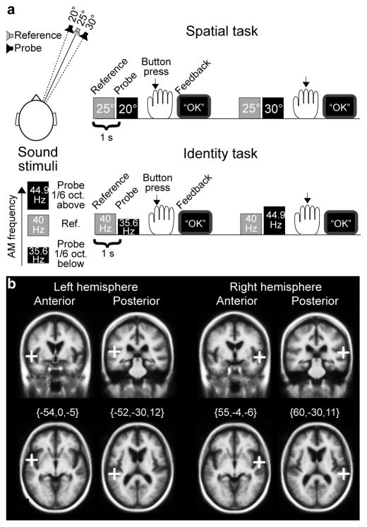
Figure 1.
Study design. (a) Stimuli and tasks. In the Spatial task, subjects discriminated whether Probe was simulated from 5° to the left or 5° to the right relative to Reference (25° to the right). In the Identity task, subjects discriminated whether Probe sound had 1/6 octaves lower or higher AM frequency than the AM frequency of Reference (40 Hz). Feedback was presented at a computer screen after each trial in both tasks. (b) Initial TMS target locations. In 50% of the trials, paired TMS pulses were delivered 55–145 ms after Probe sound onsets, bilaterally to either the posterior or anterior target regions of AC, shown here in the Freesurfer standard brain (i.e., Montreal Neurological Institute 305; MNI305) in Talairach coordinates. For each subject, target regions were transformed through the Talairach coordinate systems. Using navigated TMS, the coil was positioned to maximally stimulate the cortical area of interest. After the experiments, TMS-induced E-fields were estimated in each subject’s cortical surface using physical modeling, to localize the maximally stimulated AC subregions (Fig 4).

