
Fig 5.
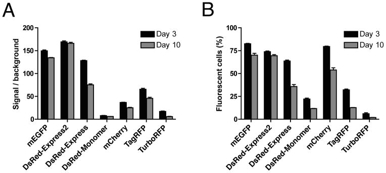
Fluorescent protein cytotoxicities after lentiviral transduction. (A) HeLa cells were transduced with lentiviral particles encoding the indicated FPs or with a control lentiviral particles lacking an FP gene. At 3 and 10 days after transduction, cells were analyzed by flow cytometry. Plotted are the average fluorescence signals from viable fluorescent cells relative to the control. (B) The percentage of viable cells that were fluorescent was also recorded at 3 and 10 days after transduction. In these experiments, DsRed-Express2 and monomeric EGFP (mEGFP, which is EGFP with the A206K mutation) maintained almost all of the original fluorescence signal and showed only a small decline in the percentage of fluorescent cells. By contrast, all of the other red FPs showed dramatic reductions in average fluorescence and in the percentage of fluorescent cells, indicating that those other FPs were cytotoxic. (Reproduced with permission from ref. 17.)