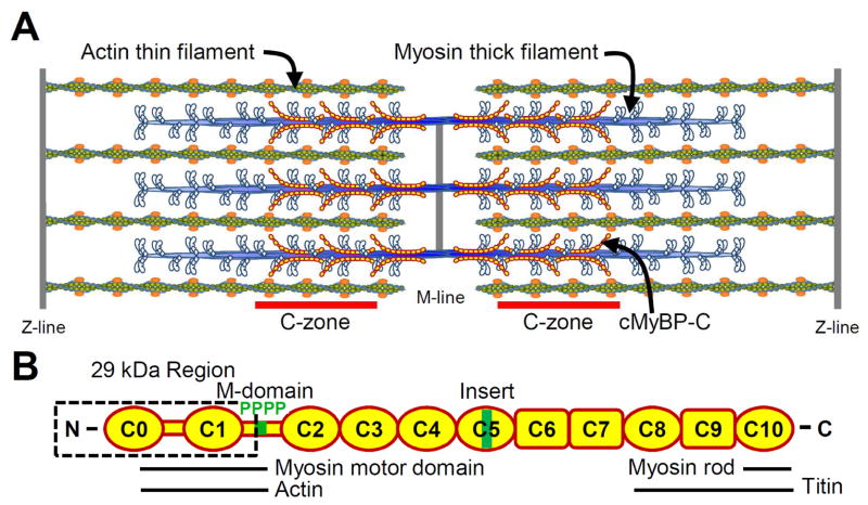
Figure 1.
A) Illustration of cardiac muscle sarcomere with interdigitating myosin-thick and actin-thin filaments. MyBP-C is localized to the C-zones. B) Schematic diagram of MyBP-C’s Ig-like (oval) and fibronectin-like (rectangle) domains structure, functionally significant 29 kDa region, M-domain with 4 phosphorylation sites and cardiac specific C5 insert. Domains involved with sarcomeric protein binding are indicated.

